The retina is the layer of tissue at the back of the eye that is responsible for vision. This tissue is attached to the choroid which supplies blood to the retina. Retinal detachment is a disease in which the retina separates from the choroid with an accumulation of liquid between these structures. This is a very serious pathology which, if not treated urgently, can lead to blindness.
Symptoms Of Retinal Detachment
There are often warning signs that can predict the onset of detachment such as flying flies, compromised vision, amputations in the visual field, and flashes of light.
Provision
Many patients have a predisposition to retinal detachment: myopia, peripheral retinal dystrophies (so-called regmatogene), familiarity with retinal detachment and operations on the ocular bulb.
Vitreous detachment
The vitreous body is a gelatinous, transparent substance that occupies the back of the eye, formed by water and hyaluronic acid. This with time after trauma, can degenerate and detach from the retina in diabetics. This phenomenon, not always pathological can be annoying or the first symptom of a retinal detachment. In most cases, the retina detaches because a hole or a retinal tear allows the liquid part of the vitreous body to pass under the retina. Most retinal detachments occur as a natural aging process in the eyes but some people are at higher risk than others. The diagnosis is made with a simple eye checkup that includes an examination of the ocular fundus.
Laser treatment
Laser treatment is used for retinal breaks without detachment or during a vitrectomy operation. The outpatient treatment lasts about 15 minutes, under local anesthesia (eye drops), with a dilated pupil. Apart from the need to use a lens on the eye, it is absolutely painless. Doctors may need to perform multiple sessions to heal a hole or dystrophy.
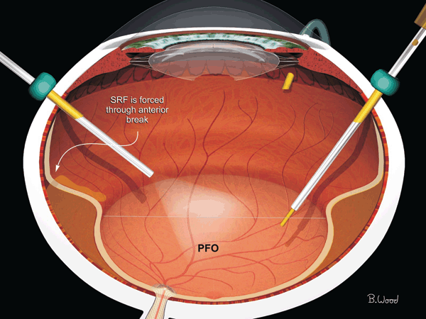
Vitrectomy: Intervention For Retinal Detachment
In most cases, the retina detaches because a hole or a retinal tear allows the liquid part of the vitreous body to pass under the retina. Most retinal detachments occur as a natural aging process in the eyes but some people are at higher risk than others. What is the treatment for retinal detachment? It requires surgery and the main goal of it is to close the holes and reattach the retina. The two methods used in retinal detachment treatment are scleral cerclage or vitrectomy or the combination of both procedures. Experienced ophthalmologists specialized in this field perform this very complex operation. When they plan an ab-externo treatment (scleral cerclage), they use a silicone band. The band creates a ridge inside the eye and pushes the outer wall of the eye against the hole in the retina.
The silicone band is not visible on the outer wall of the eye and usually remains in place permanently. The intervention lasts about 30 minutes even if the intervention times are different from case to case, linked to the pathology. The operation is performed under local anesthesia with the assistance of an anesthesiologist. In some cases, general anesthesia may be used.
Vitrectomy
To heal the retinal break you can use the laser or a probe in cold temperature. With vitrectomy air, a gas bubble or silicone oil is used to help the retina in the healing process. The gas bubble is slowly absorbed in about 2-6 weeks while the silicone oil needs a second operation for removal after 3-6 months. With the use of these buffering systems (air, gas, silicone) the view will be very blurred and inconsistent over time. With these substances in the postoperative period, a postural positioning will be necessary to favor the repositioning of the retina.
Postural Positioning After Surgery
When the eye is injected with these buffering substances (air, gas, silicone oil) it may be necessary to position the head and eye in such a way that it helps the retina to be relocated. There are various postural positions and the surgeon will recommend the most suitable one. The posture is often the most difficult part of recovery after surgery. But, it is very important and should be considered as the second phase of the operation.
Risks of Retinal Detachment Surgery
Interventions for retinal detachment (circling and/or vitrectomy) are sometimes not successful during the first operation. Hence, there may be a need for other interventions to take place in order to achieve the repositioning of the retina. The rate of success is about 80% for retinal detachment surgery with a single operation. This means that 2 out of 10 people (10%) will need more than one operation. If a gas or oil bubble is used during surgery it will develop a cataract in the eye within two years. Any surgical intervention carries a risk of bleeding and infections but in retinal detachment surgery, this risk is very low (less than one in a thousand). However, if it does occur, it can have serious consequences until it causes blindness.
The Postoperative Period
Although the retinal detachment surgery is an invasive operation, it is rarely painful and normal postoperative discomforts can be treated with systemic painkillers. The redness of the conjunctiva & sclera (white of the eye) and swollen eyelids are normal in the following fifteen days. These symptoms gradually disappear with local and general therapy. The sight in the operated eye will be quite blurry for the first few weeks. The recovery of sight will be gradual and can take several weeks or months. The patient may need new glasses. It is important to consult the surgeon and check the postoperative status.
Vitrectomy Recovery Time
The recovery of the visus after a retinal detachment intervention can be slow and tedious. In many cases, sight recovery is incomplete due to macula involvement, the central zone of vision. Patients often need to update their glasses prescription afterward.
Work Activity After The Intervention
Most patients take about fifteen days off work. The exact time depends on the type of work, surgery, and retinal detachment. In some cases, the postoperative period may be longer.
Subsequent Interventions
The retinal detachment can recur in 20-30%. In these cases, a new intervention is necessary generally for a vitrectomy operation. Recurrences occur mainly due to the onset of PVR (Vitreo-Retinal Proliferation). It is an important cicatricial reaction with the formation of fibrous membranes and tractions that determine a new retinal detachment. This condition is treated with a vitrectomy, during which silicone oil is introduced and later removed.
The Intervention of Removing Silicone Oil
Silicone is removed once it has served its purpose of reattaching the retina, usually after 3–6 months. Factors that can speed up or delay silicone removal include eye pressure, cataracts, retinal detachment recurrence, vitreoretinal proliferation, emulsification, and silicone bubble dislocation.
The silicone oil removal procedure can vary by case, including atrophic holes, macular holes, or partial retinal detachment recurrences. In conclusion, the decision to remove silicone must be personalized for each patient.